
Electron Microscopy
To see what no one has seen before
Whether you are interested in learning how to do electron microscopy (EM) yourself or prefer us to do EM for you – we can help! We offer a wide variety of preparation and visualization techniques for biological samples ranging from standard methods to cutting-edge cryo-EM for high-resolution 2D or 3D imaging. We also provide a basic scanning EM service to visualize surface structures of biological samples.
Need EM of a biological sample? Want to learn how to do EM yourself? Interested in learning more about our EM services?

Electron microscopy training and infrastructure usage
Infrastructure users receive thorough training in the techniques/instruments they are interested in. All our instruments - from basic sample preparation equipment to our most sophisticated electron microscopes - are available to users 24/7. We further support our users in planning, execution, and interpretation of all EM-related experiments and regularly organize EM workshops and microscopy training.


EM service to visualize biological samples
We can do EM for you and have experience in the visualization of the following samples:
- Molecules (RNA, DNA, proteins, lipids)
- Complexes (liposomes, emulsions, synthetic structures)
- Viruses
- Bacteria
- Organelles
- Eukaryotic cells (fungi, plants, animals)
- Tissues
- Surface structures
We are also very interested in establishing protocols for new samples.

Sample preparation techniques
We offer a wide variety of different sample preparation techniques to ensure optimal preservation of your specimen:
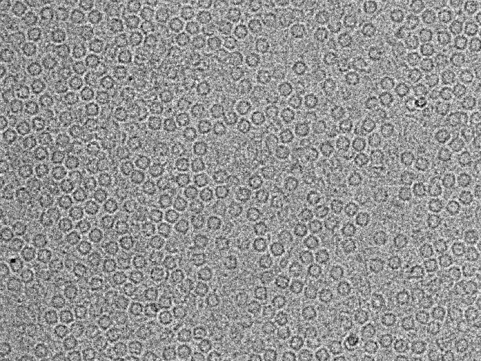
- Negative staining for rapid visualization of macromolecules, proteins, viruses, organelles, or bacteria
- Conventional chemical fixation for tissue samples and cell monolayers
- High pressure freezing and freeze substitution for superior cellular preservation of cells and tissues
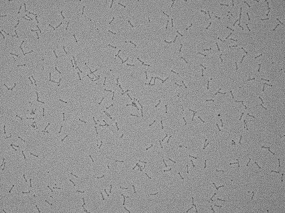
- Rotary shadowing for high contrast examination of protein complexes
- Freeze fracturing and etching for visualization of surfaces, membranes, and the inside of membranous compartments
- Plunge freezing (immersion freezing) for cryo-EM (“cryo plunge freezing”) for best possible preservation of small samples close to their native state
- Tokuyasu as a method for cryo-sectioning in combination with immunogold labeling
- Immunolabeling to locate structures of interest
- Ultramicrotomy: ultra-thin sectioning of resin-embedded samples
- Sample preparation for SEM (scanning electron microscopy) to study surface structures

Imaging techniques
We offer the following EM imaging techniques to visualize your samples:
- Basic Scanning Electron Microscopy (SEM) to visualize the surface topology of samples
- Transmission Electron Microscopy (TEM) to examine ultrastructure
- Conventional 2D EM
- Tomography - 3D EM
- Cryo-EM (single particle and cryo-electron tomography)
- Correlative Microscopy


EQUIPMENT
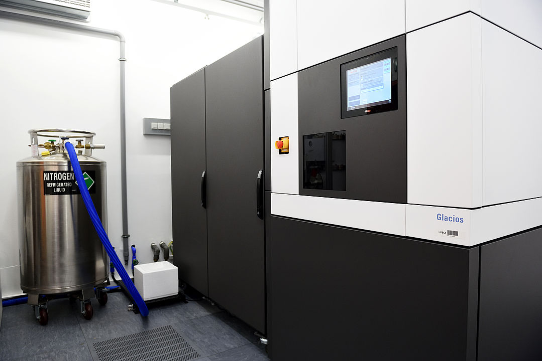
The 200 kV Thermo Fisher Scientific Glacios is a cryo-TEM for high-throughput sample screening and fully automated data recording. The Glacios is exclusively used for cryo-EM (single particle and cryo-tomography). Watch a time-lapse video of the installation below.
Please click here for more information.
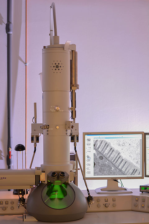
The FEI Morgagni 268D is a robust and easy-to-use 100 kV TEM equipped with an Mega View III CCD (Olympus-SIS) camera. The microscope is easy to use and tailored for sample screening and routine visualization of:
- Negatively-stained samples
- Rotary-shadowed samples
- Ultrathin sections
- Immunolabelled samples
Please click here for more information.
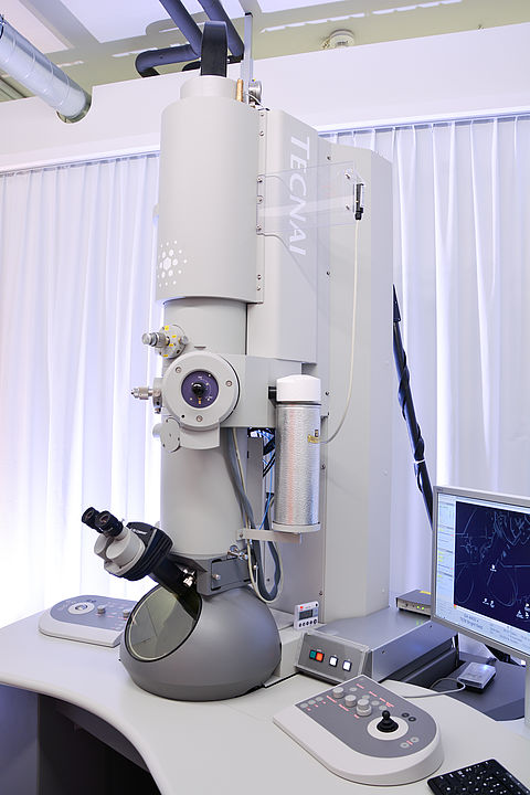
The FEI Tecnai G2 20 (T20) is a 200 kV TEM equipped with an Eagle 4k HS camera and can be used for 2D and 3D (tomography) visualization of:
- Negatively-stained samples (screening and automated data recording)
- Rotary-shadowed samples
- Room temperature tomography of resin-embedded samples
Please click here for more information.
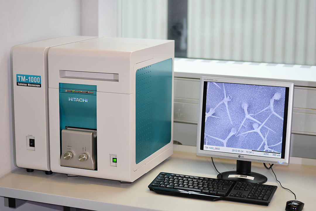
This is a robust and easy-to-use table top scanning electron microscope for visualization of surface properties at higher resolution and deeper focus depth than achievable by light microscopy. The image is generated with back-scattered electrons induced by an electron beam of 15 keV.
Sample preparation is straightforward since it does not require coating with a conductive layer. The biological material can be frozen in liquid nitrogen. Alternatively, it can be chemically fixed and critical point-dried.

Additionally, we offer the following equipment and techniques:
- Immersion and slam freezing
- High vacuum evaporators and sputter coaters
- High pressure freezing and freeze substitution
- Sectioning
- Embedding
- Sample preparation for scanning electron microscopy
- Ancillary equipment
- Light microscopes
Please click here for more information.
Installation of GLACIOS at VBCF Electron Microscopy Facility

USER INFORMATION
In general, we offer four types of access to our shared research infrastructure:
- Research projects
- Full research services
- Trained user access | user labs
- Shared technology platform
Typically, these are set but not limited by the offered technology or instrument, and differ in the required user expertise, the usability of a technology, the user’s pre- and postprocessing input, and the underlying operational models.
Research project
Research projects are the equivalent of contract research organizations (CRO). The customer submits the starting material/sample and receives the ready-to-use data for publication. Hence, core facility members are often co-authoring and involved in the entire publication process.
Full research service
The user submits the sample, we perform a pre-defined workflow (incl. QC) and process the raw data. Data interpretation or contributions to publications cannot be offered in this service mode.
Trained user access | User labs
VBCF experts maintain an instrument park and train users to operate it. This requires a certain level of expertise, maturity of the offered technology, a hands-on attitude and reliability from the user.
Shared technology platform | Instrument park
These technologies require expert knowledge to run the offered instruments. Experts are hired by one of the research institutes on the Vienna BioCenter Campus. The experts maintain an instrument park, run the experiments and train other trainers. Machines can only be operated by experts.
The VBCF Electron Microscopy facility can be accessed via research projects. The facility also provides full research service and trained user access.
In general, the VBCF General Cooperation Conditions apply. If you want to use equipment or require training, please fill out the Infrastructure Usage and Training Request form. To book your services, please provide the following details in a service request:
- What is your scientific question?
- Which organisms and samples are you working with?
- Do you have preliminary results?
- Are there any papers describing what you want to do?
We require acknowledgement of facility use in publications. A simple statement is sufficient and can be placed in the Materials and Methods section or in the Acknowledgments section, depending on the journal format.
Suggested format:
The XXXXXX was performed by the Electron Microscopy Facility at Vienna BioCenter Core Facilities (VBCF), member of the Vienna BioCenter (VBC), Austria.
In case of (co-)authorship:
The Vienna BioCenter Core Facilities (VBCF) Electron Microscopy Facility acknowledges funding from the Austrian Federal Ministry of Education, Science & Research; and the City of Vienna.
Collaborating Labs
- CIUS Vienna - Cell Imaging and Ultrastructure Research
- ZMF Graz - Core Facility Ultrastructure Analysis
- USTEM TU Vienna - Service Facility for Transmission Electron Microscopy
- Nexperion
- IST Austria
EM Instrumentation Manufacturers
Software Tools
Electron Microscopy Journals
- Journal of Microscopy
- Journal of Structural Biology
- Ultramicroscopy
- Micron
- Journal of Electron Microscopy
Discussion Groups and Mailing Lists
Societies
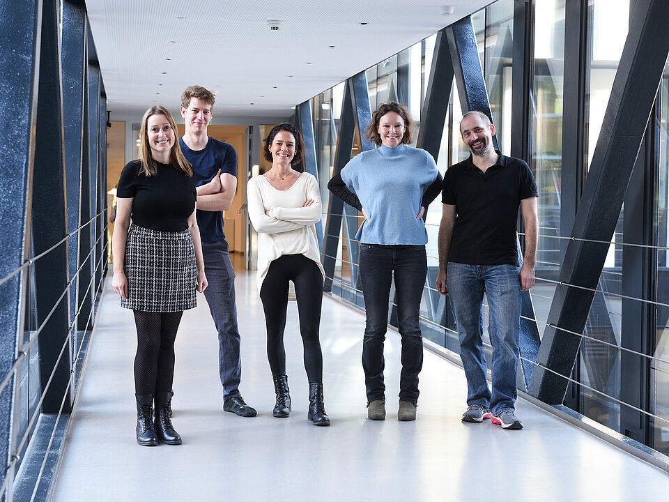
VBCF ELECTRON MICROSCOPY TEAM
SCIENTIFIC CONTRIBUTIONS
Analysis of extracellular vesicle microRNA profiles reveals distinct blood and lymphatic endothelial cell origins. Pultar M, Oesterreicher J, Hartmann J, Weigl M, Diendorfer A, Schimek K, Schädl B, Heuser T, Brandstetter M, Grillari J, Sykacek P, Hackl M, Holnthoner W. (2024) Journal of Extracellular Biology Vol3, Issue 1, January 2024, e134
A rapid freezing method to determine tissue layer thickness in drought-stressed leaves. Zekri MA, Leimhofer C, Drexler N, Lang I (2024) Journal of Microscopy, 2024; 1-9.
Structural basis of human U5 snRNP late biogenesis and recycling. Riabov Bassat D, Visanpattanasin S, Vorländer M.K., Fin L, Phillips AW, Plaschka C (2024) Nat Struct Mol Biol (2024).
Generation of complex bone marrow organoids from human induced pluripotent stem cells. Frenz-Wiessner S, Fairley SD, Buser M, Goek I, Salewskij K, Jonsson G, Illig D, Putlitz B, Petersheim D, Li Y, Chen P, Kalauz M, Conca R, Sterr M, Geuder J, Mizoguchi Y, Megens RTA, Linder MI, Kotlarz D, Rudelius M, Penninger JM, Marr C, Klein C. Nat Methods (2024).
Morphological and molecular characterization of Stomachicola muraenesocis Yamaguti, 1934 (Digenea: Hemiuridae) from the daggertooth pike conger Muraenesox cinereus (Forsskål). Ghanei-Motlagh R., Hernández-Orts J.S., Fast M.D., Whyte S.K., El-Matbouli M., Saleh M. Parasitology. 2023:1-21.
Adeno-Associated Virus-like Particles' Response to pH Changes as Revealed by nES-DMA. Zoratto S, Heuser T, Friedbacher G, Pletzenauer R, Graninger M, Marchetti-Deschmann M, Weiss VU. Viruses. 2023 Jun 13;15(6):1361.
Time-resolved cryo-EM (TR-EM) analysis of substrate polyubiquitination by the RING E3 anaphase-promoting complex/cyclosome (APC/C). Bodrug T, Welsh KA, Bolhuis DL, Paulаkonis E, Martinez-Chacin RC, Liu B, Pinkin N, Bonacci T, Cui L, Xu P, Roscow O, Amann SJ, Grishkovskaya I, Emanuele MJ, Harrison JS, Steimel JP, Hahn KM, Zhang W, Zhong ED, Haselbach D, Brown NG. Nat Struct Mol Biol. 2023 Nov;30(11):1663-1674.
Structural evidence for elastic tethers connecting separating chromosomes in crane-fly spermatocytes. Forer A, Otsuka S. Life Sci Alliance. 2023 Aug 17;6(11):e202302303.
Human serum albumin nanoparticles as a versatile vehicle for targeted delivery of antibiotics to combat bacterial infections. Skoll K, Palmetzhofer J, Lummerstorfer M, Anzengruber M, Gabor F, Wirth M. Nanomedicine: Nanotechnology, Biology and Medicine, Volume 50, 2023, 102685, ISSN 1549-9634.
A phylogenetic profiling approach identifies novel ciliogenesis genes in Drosophila and C. elegans. Dobbelaere J., Su Y. T., Erdi B., Schleiffer A., Dammermann A. The EMBO Journal 2023 e113616.
Cryo-EM structure of the chain-elongating E3 ubiquitin ligase UBR5. Hodáková Z., Grishkovskaya I., Brunner L. H., Bolhuis D.L, Belačić K, Schleiffer A., Kotisch H., Brown N.B., Haselbach D. The EMBO Journal 2023 42:e113348.
Archaeosomes facilitate storage and oral delivery of cannabidiol. Sedlmayr V., Horn C., Wurm D.J., Spadiut O., Quehenberger J. International Journal of Pharmaceutics 2023, Volume 645, 2023, 123434, ISSN 0378-5173.
Lipid saturation controls nuclear envelope function. Romanauska A. & Köhler A. Nat Cell Biol 25, 1290–1302 2023.
Morphological Study of PHA Producing Bacteria. Hrubanova K., Sikorova P., Mrázová K., Nebesarova J., Obruca S., Krzyzanek V. Microscopy and Microanalysis, Volume 29, Issue Supplement_1, 1 August 2023, Pages 883–884.
Structure and regulation of the myotonic dystrophy kinase-related Cdc42-binding kinase. Truebestein L, Antonioli S, Waltenberger E, Gehin C, Gavin AC, Leonard TA. Structure, 31,435-446.e4.
Description, molecular identification and pathological lesions of Huffmanela persica sp. nov. (Nematoda: Trichosomoididae: Huffmanelinae) from the daggertooth pike conger Muraenesox cinereus. Ghanei-Motlagh R, Fast MD, Groman D, Kumar G, Soliman H, El-Matbouli M, Saleh M. Parasites Vectors 16, 182 (2023).
Structural basis for regulation of apoptosis and autophagy by the BIRC6/SMAC complex. Ehrmann JF, Grabarczyk DB, Heinke M, Deszcz L, Kurzbauer R, Hudecz O, Shulkina A, Gogova R, Meinhart A, Versteeg GA, Clausen T. Science. 2023 379(6637):1117-1123.
Extracellular Vesicles and Particles Modulate Proton Secretion in a Model of Human Parietal Cells. Mistlberger-Reiner A, Sterneder S, Reipert S, Wolske S, Somoza V. ACS Omega. 2023 8(2):2213-2226.
Characterization of membrane vesicles in Alteromonas macleodii indicates potential roles in their copiotrophic lifestyle, Fadeev E, Carpaneto Bastos C, Hennenfeind JH, Biller SJ, Sher D, Wietz M, Herndl GJ, microLife 2023, 4, uqac025
mRNA recognition and packaging by the human transcription-export complex. Pacheco-Fiallos B, Vorländer MK, Riabov-Bassat D, Fin L, O'Reilly FJ, Ayala FI, Schellhaas U, Rappsilber J, Plaschka C. Nature. 2023 Apr 5. doi: 10.1038/s41586-023-05904-0. Epub ahead of print.
Aerosol delivered irradiated Escherichia coli confers serotype-independent protection and prevents colibacillosis in young chickens. Paudel S, Hess C, Kamal Abdelhamid M, Lyrakis M, Wijewardana V, Thiga Kangethe R, Cattoli G, Hess M. Vaccine. 2023 41(7):1342-1353.
A molecular network of conserved factors keeps ribosomes dormant in the egg. Leesch F, Lorenzo-Orts L, Pribitzer C, Grishkovskaya I, Roehsner J, Chugunova A, Matzinger M, Roitinger E, Belačić K, Kandolf S, Lin TY, Mechtler K, Meinhart A, Haselbach D, Pauli A. Nature. 2023 613(7945):712-720
Stabilization of the Quadruplex-Forming G-Rich Sequences in the Rhinovirus Genome Inhibits Uncoating—Role of Na+ and K+. Real-Hohn A, Groznica M, Kontaxis G, Zhu R, Chaves OA, Vazquez L, Hinterdorfer P, Kowalski H, Blaas D Viruses. 2023; 15(4):1003.
PCYT2-regulated lipid biosynthesis is critical to muscle health and ageing. Cikes D, Elsayad K, Sezgin E, Koitai E, Torma F, Orthofer M, Yarwood R, Heinz LX, Sedlyarov V, Miranda ND, Taylor A, Grapentine S, Al-Murshedi F, Abot A, Weidinger A, Kutchukian C, Sanchez C, Cronin SJF, Novatchkova M, Kavirayani A, Schuetz T, Haubner B, Haas L, Hagelkruys A, Jackowski S, Kozlov AV, Jacquemond V, Knauf C, Superti-Furga G, Rullman E, Gustafsson T, McDermot J, Lowe M, Radak Z, Chamberlain JS, Bakovic M, Banka S, Penninger JM. Nat Metab. 2023 5(3):495-515.
Generation of Human Induced Pluripotent Stem Cell-Derived Bone Marrow Organoids. Frenz S, Goeki I, Buser M, Salewskij K, Fairley S, Conca R, Drexler N, Jonsson G, Thomas M, Mizoguchi Y, Rudelius M, Heuser T, Marr C, Penninger JM, Klein C. Blood (2022) 140 (Supplement 1): 1682–1683.
The Xbp1-regulated transcription factor Mist1 restricts antibody secretion by restraining Blimp1 expression in plasma cells. Wöhner M, Pinter T, Bönelt P, Hagelkruys A, Kostanova-Poliakova D, Stadlmann J, Konieczny SF, Fischer M, Jaritz M, Busslinger M. Front Immunol. 2022 13:859598.
EVAnalyzer: High content imaging for rigorous characterisation of single extracellular vesicles using standard laboratory equipment and a new open-source ImageJ/Fiji plugin. Schürz M, Danmayr J, Jaritsch M, Klinglmayr E, Benirschke HM, Matea CT, Zimmerebner P, Rauter J, Wolf M, Gomes FG, Kratochvil Z, Heger Z, Miller A, Heuser T, Stanojlovic V, Kiefer J, Plank T, Johnson L, Himly M, Blöchl C, Huber CG, Hintersteiner M, Meisner-Kober N. J Extracell Vesicles. 2022 11(12):e12282.
Structure of the peripheral arm of a minimalistic respiratory complex I. Schimpf J, Oppermann S, Gerasimova T, Santos Seica AF, Hellwig P, Grishkovskaya I, Wohlwend D, Haselbach D, Friedrich T. Structure. 2022 30(1):80-94.e4
Transcriptome, metabolome and suppressor analysis reveal an essential role for the ubiquitin-proteasome system in seedling chloroplast development. Talloji P, Nehlin L, Hüttel B, Winter N, Černý M, Dufková H, Hamali B, Hanczaryk K, Novák J, Hermanns M, Drexler N, Eifler K, Schlaich N, Brzobohatý B, Bachmair A. BMC Plant Biol. 2022 22(1):183.
p57Kip2 imposes the reserve stem cell state of gastric chief cells. Lee JH, Kim S, Han S, Min J, Caldwell B, Bamford AD, Rocha ASB, Park J, Lee S, Wu SS, Lee H, Fink J, Pilat-Carotta S, Kim J, Josserand M, Szep-Bakonyi R, An Y, Ju YS, Philpott A, Simons BD, Stange DE, Choi E, Koo BK, Kim JK. Cell Stem Cell. 2022 29(5):826-839.e9.
Introduction to cryo-transmission electron microscopy.Hielkema L., Heuser T., Paulino C. and Walter A.; 2022 Chapter 2; This chapter was published in the textbook Imaging Modalities For Biological And Preclinical Research: A compendium, Volume 1 (IOP Publishing Ltd,2021) ISBN: 978-0-7503-3059-6
A functional corona around extracellular vesicles enhances angiogenesis, skin regeneration and immunomodulation. Wolf M, Poupardin RW, Ebner-Peking P, Andrade AC, Blöchl C, Obermayer A, Gomes FG, Vari B, Maeding N, Eminger E, Binder HM, Raninger AM, Hochmann S, Brachtl G, Spittler A, Heuser T, Ofir R, Huber CG, Aberman Z, Schallmoser K, Volk HD, Strunk D. J Extracell Vesicles. 2022 11(4):e12207.
Release of CHK-2 from PPM-1.D anchorage schedules meiotic entry. Baudrimont A, Paouneskou D, Mohammad A, Lichtenberger R, Blundon J, Kim Y, Hartl M, Falk S, Schedl T, Jantsch V. Sci Adv. 2022 8(7):eabl8861.
Cryo-EM structure of the plant 26S proteasome. Kandolf S, Grishkovskaya I, Belačić K, Bolhuis DL, Amann S, Foster B, Imre R, Mechtler K, Schleiffer A, Tagare HD, Zhong ED, Meinhart A, Brown NG, Haselbach D. Plant Commun. 2022 3(3):100310.
BacPROTACs mediate targeted protein degradation in bacteria. Morreale FE, Kleine S, Leodolter J, Junker S, Hoi DM, Ovchinnikov S, Okun A, Kley J, Kurzbauer R, Junk L, Guha S, Podlesainski D, Kazmaier U, Boehmelt G, Weinstabl H, Rumpel K, Schmiedel VM, Hartl M, Haselbach D, Meinhart A, Kaiser M, Clausen T. Cell. 2022 185(13):2338-2353.e18.
Lysosomal enzyme trafficking factor LYSET enables nutritional usage of extracellular proteins. Pechincha C, Groessl S, Kalis R, de Almeida M, Zanotti A, Wittmann M, Schneider M, de Campos RP, Rieser S, Brandstetter M, Schleiffer A, Müller-Decker K, Helm D, Jabs S, Haselbach D, Lemberg MK, Zuber J, Palm W. Science. 2022: eabn5637
A mitotic chromatin phase transition prevents perforation by microtubules. Schneider MWG, Gibson BA, Otsuka S, Spicer MFD, Petrovic M, Blaukopf C, Langer CCH, Batty P, Nagaraju T, Doolittle LK, Rosen MK, Gerlich DW. Nature. 2022 609(7925):183-190.
Transverse Electron-Beam Shaping with Light. Chirita Mihaila MC, Weber P, Schneller M, Grandits L, Nimmrichter S, Juffmann T Phys. Rev. X 2022 12, 031043
AKIRIN2 controls the nuclear import of proteasomes in vertebrates. de Almeida M, Hinterndorfer M, Brunner H, Grishkovskaya I, Singh K, Schleiffer A, Jude J, Deswal S, Kalis R, Vunjak M, Lendl T, Imre R, Roitinger E, Neumann T, Kandolf S, Schutzbier M, Mechtler K, Versteeg GA, Haselbach D, Zuber J. Nature. 2021 599(7885):491-496.
Scalable Enrichment of Immunomodulatory Human Acute Myeloid Leukemia Cell Line-Derived Extracellular Vesicles. Binder HM, Maeding N, Wolf M, Cronemberger Andrade A, Vari B, Krisch L, Gomes FG, Blöchl C, Muigg K, Poupardin R, Raninger AM, Heuser T, Obermayer A, Ebner-Peking P, Pleyer L, Greil R, Huber CG, Schallmoser K, Strunk D. Cells. 2021 10(12):3321.
miR-1 sustains muscle physiology by controlling V-ATPase complex assembly. Gutiérrez-Pérez P, Santillán EM, Lendl T, Wang J, Schrempf A, Steinacker TL, Asparuhova M, Brandstetter M, Haselbach D, Cochella L. Sci Adv. 2021 7(42):eabh1434.
Cohesin mediates DNA loop extrusion by a "swing and clamp" mechanism. Bauer BW, Davidson IF, Canena D, Wutz G, Tang W, Litos G, Horn S, Hinterdorfer P, Peters JM. Cell. 2021 184(21):5448-5464.e22.
Glycoproteins of Predicted Amphibian and Reptile Lyssaviruses Can Mediate Infection of Mammalian and Reptile Cells. Oberhuber M, Schopf A, Hennrich AA, Santos-Mandujano R, Huhn AG, Seitz S, Riedel C, Conzelmann KK. Viruses. 2021 13(9):1726.
An inhibitor-mediated beta cell dedifferentiation model reveals distinct roles for FoxO1 in glucagon repression and insulin maturation. Casteels T, Zhang Y, Frogne T, Sturtzel C, Lardeau CH, Sen I, Liu X, Hong S, Pauler FM, Penz T, Brandstetter M, Barbieux C, Berishvili E, Heuser T, Bock C, Riedel CG, Meyer D, Distel M, Hecksher-Sørensen J, Li J, Kubicek S. Mol Metab. 2021 101329
Reprogrammed lipid metabolism protects inner nuclear membrane against unsaturated fat. Romanauska A, Köhler A. Dev Cell. 2021 56(18):2562-2578.e3.
HUWE1 employs a giant substrate-binding ring to feed and regulate its HECT E3 domain. Grabarczyk DB, Petrova OA, Deszcz L, Kurzbauer R, Murphy P, Ahel J, Vogel A, Gogova R, Faas V, Kordic D, Schleiffer A, Meinhart A, Imre R, Lehner A, Neuhold J, Bader G, Stolt-Bergner P, Böttcher J, Wolkerstorfer B, Fischer G, Grishkovskaya I, Haselbach D, Kessler D, Clausen T. Nat Chem Biol. 2021 17(10):1084-1092.
Cross-modality imaging of bisphosphonate-treated murine jawbones. Reier S, Turyanskaya A, Heimel P, Frischauf N, Meusburger D, Heuser T, Drexler N, Janovszky Á, Streli C, Slezak P, Plochberger B, Dungel P, Szabó A, Walter A. Analyst. 2021 146(14):4683-4699.
Effect of hydroxypropyl‐β‐cyclodextrin in fluid and semi‐solid submicron emulsions on physiological skin parameters during regular in vivo application. Pany A., Wohlgenannt M., Klopprogge S., Wolzt M., Heuser T., Kotisch H., Valenta C., Klang V. (2021) Int J Cosmet Sci, 43(2):263-268
Binding Mode Characterization of Osteopontin on Hydroxyapatite by Solution NMR Spectroscopy. Holzinger J, Kotisch H, Richter KW, Konrat R (2021) ChemBioChem 2021, 22, 1-7.
Substrate-engaged type III secretion system structures reveal gating mechanism for unfolded protein translocation. Miletic S, Fahrenkamp D, Goessweiner-Mohr N, Wald J, Pantel M, Vesper O, Kotov V, Marlovits TC. Nat Commun. 2021 12(1):1546.
The linear ubiquitin chain assembly complex (LUBAC) generates heterotypic ubiquitin chains. Rodriguez Carvajal A, Grishkovskaya I, Gomez Diaz C, Vogel A, Sonn-Segev A, Kushwah MS, Schodl K, Deszcz L, Orban-Nemeth Z, Sakamoto S, Mechtler K, Kukura P, Clausen T, Haselbach D, Ikeda F. Elife. 2021 10:e60660.
Structural basis for inhibition of the AAA-ATPase Drg1 by diazaborine. Prattes M, Grishkovskaya I, Hodirnau VV, Rössler I, Klein I, Hetzmannseder C, Zisser G, Gruber CC, Gruber K, Haselbach D, Bergler H. Nat Commun. 2021 12(1):3483.
The dimeric Golgi protein Gorab binds to Sas6 as a monomer to mediate centriole duplication. Fatalska A, Stepinac E, Richter M, Kovacs L, Pietras Z, Puchinger M, Dong G, Dadlez M, Glover DM. Elife. 2021 10:e57241
Cryo-EM grid optimization for membrane proteins. Kampjut D, Steiner J and Sazanov L (2021) iScience 24(3):102139.
An acentriolar centrosome at the C. elegans ciliary base. Garbrecht J, Laos T, Holzer E, Dillinger M and Dammermann A (2021) Current Biology 31(11):2418-2428.e8.
A crucial role for Jagunal homolog 1 in humoral immunity and antibody glycosylation in mice and humans. Hagelkruys A, Wirnsberger G, Stadlmann J, Wöhner M, Horrer M, Vilagos B, Jonsson G, Kogler M, Tortola L, Novatchkova M, Bönelt P, Hoffmann D, Koglgruber R, Steffen U, Schett G, Busslinger M, Bergthaler A, Klein C, Penninger JM (2021) J Exp Med. 218(1):e20200559.
Safe and effective two-in-one replicon-and-VLP minispike vaccine for COVID-19: Protection of mice after a single immunization.Hennrich AA,Sawatsky B,Santos-Mandujano R,Banda DH,Oberhuber M,Schopf A,Pfaffinger V, Wittwer K,Riedel C,Pfaller CK, Conzelmann K (2021) PLoS Pathog 17(4): e1009064.
Cardioids reveal self-organizing principles of human cardiogenesis. Hofbauer P, Jahnel SM, Papai N, Giesshammer M, Deyett A, Schmidt C, Penc M, Tavernini K, Grdseloff N, Meledeth C, Ginistrelli LC, Ctortecka C, Salic S, Novatchkova M, Mendjan S (2021) Cell 184 (12) 3299-3317.e22.
Components and Architecture of the Rhabdovirus Ribonucleoprotein Complex. Riedel C, Hennrich AA, Conzelmann KK. Viruses. 2020 12(9):959.
Cell death induced autophagy contributes to terminal differentiation of skin and skin appendages. Koenig U., Robenek H., Barresi C., Brandstetter M., Resch GP., Gröger M, Pap T., Hartmann C. (2020) Autophagy 16(5):932-945
Structures of three MORN repeat proteins and a re- evaluation of the proposed lipid-binding properties of MORN repeats. Sajko S, Grishkovskaya I, Kostan J, Graewert M, Setiawan K, Trübestein L, Niedermüller K, Gehin C, Sponga A, Puchinger M, Gavin A, Leonard T, Svergun I, Smith T, Morriswood B, Djinovic-Carugo K (2020) PLoS ONE 15(12): e0242677
Repression of the B cell identity factor Pax5 is not required for plasma cell development. Grace Liu J., Jaritz M., Wöhner M., Agerer B., Bergthaler A., Malin S.G., and Busslinger M. (2020) J. Exp. Med. 2020 Vol. 217 No. 11 e20200147.
The Brownian and Flow‐Driven Rotational Dynamics of a Multicomponent DNA Origami‐Based Rotor. Ahmadi Y., Nord A. L., Wilson A. J., Hütter C., Schroeder F., Beeby M., Barisic I. (2020) Small/Vol. 16, Issue 22.
Vaccinia Virus Immunomodulator A46: Destructive Interactions with MAL and MyD88 Shown by Negative-Stain Electron Microscopy. Azar D.F., Haas M., Fedosyuk S., Rahaman Md. H., Hedger A., Kobe B., Skern T. (2020) J.Structure.2020.09.007
Quantifying the heterogeneity of macromolecular machines by mass photometry. Sonn-Segev A., Belacic K., Bodrug T., Young G., VanderLinden R.T., Schulman B. A., Schimpf J., Friedrich T., Dip P.V., Schwartz T.U., Bauer B., Peters J.M., Struwe W.B., Benesch J.L.P., Brown N.G., Haselbach D. & Kukura P. (2020) Nature Communications 11, 1772 (2020)
Monomeric and homotrimeric solution structrures of truncated human peroxidasin 1 variants. Paumann-Page M., Tscheliessnig R., Sevcnikar B., Katz R., Schwartz I., Hofbauer S., Pfanzagl V., Furtmüller P. G., Obinger C. (2020) BBA - Proteins and Proteomics 1868 (2020) 140249
3D printed cell culture grid holders for improved cellular specimen preparation in cryo-electron microscopy. Fäßler F., Zens B., Hauschild R., Schur F.K.M. (2020) J Struct Biol. Vol.212, Issue 3, 1 December 2020, 107633
Moyamoya disease factor RNF213 is a giant E3 ligase with a dynein-like core and a distinct ubiquitin-transfer mechanism. Ahel J., Lehner A., Vogel A., Schleiffer A., Meinhart A., Haselbach D. and Clausen T. eLife 9: e56185
A cross-kingdom conserved ER-phagy receptor maintains endoplasmic reticulum homeostasis during stress. Stephani M, Picchianti L, Gajic A, Beveridge R, Skarwan E, Sanchez de Medina Hernandez V, Mohseni A, Clavel M, Zeng Y, Naumann C, Matuszkiewicz M, Turco E, Loefke C, Li B, Dürnberger G, Schutzbier M, Chen HT, Abdrakhmanov A, Savova A, Chia KS, Djamei A, Schaffner I, Abel S, Jiang L, Mechtler K, Ikeda F, Martens S, Clausen T, Dagdas Y. [2020] Elife 9:e58396.
Oxidative Metabolism Drives Immortalization of Neural Stem Cells during Tumorigenesis Bonnay F, Veloso A, Steinmann V, Köcher T, Abdusselamoglu MD, Bajaj S, Rivelles E, Landskron L, Esterbauer H, Zinzen RP, Knoblich JA. [2020] Cell 182 (6), 1490-1507
Calcium modulates the domain flexibility and function of an α-actinin similar to the ancestral α-actinin. Pinotsis N, Zielinska K, Babuta M, Arolas JL, Kostan J, Khan MB, Schreiner C, Salmazo A, Ciccarelli L, Puchinger M,Gkougkoulia EA, Ribeiro E, Marlovits TC, Bhattacharya A, Djinovic-Carugo K [2020] PNAS 117 (36) 22101-22112
Structure of the human core transcription-export complex reveals a hub for multivalent interactions.Pühringer T, Hohmann U, Fin L, Pacheco-Fiallos B, Schellhaas U, Brennecke J, Plaschka C [2020] eLife 2020;9:e61503
A human tissue screen identifies a regulator of ER secretion as a brain-size determinant. Esk C, Lindenhofer D, Haendeler S,Wester RA, Pflug F, Schroeder B, Bagley JA, Elling U, Zuber J, von Haeseler A, Knoblich JA [2020] Science 370, 6519, pp. 935-941
Cep97 Is Required for Centriole Structural Integrity and Cilia Formation in Drosophila. Dobbelaere J, Schmidt Cernohorska M, Huranova M, Slade D, Dammermann A [2020] Current Biology 30,15, 3045-3056.e7.
A crucial role for Jagunal homolog 1 in humoral immunity and antibody glycosylation in mice and humans. Hagelkruys A, Wirnsberger G, Stadlmann J, et al. [2020] J Exp Med 218 (1): e20200559
Fractal dimension of antibody‐PEG precipitate: Light microscopy for the reconstruction of 3D precipitate structures. Satzer, P, Burgstaller, D, Krepper, W, Jungbauer, A [2020] Eng Life Sci. 20: 67– 78.
Short-course radiotherapy promotes pro-inflammatory macrophages via extracellular vesicles in human rectal cancer. Stary V, Wolf B, Unterleuthner D, et al [2020] Journal for ImmunoTherapy of Cancer 8:e000667.
Reconstitution of autophagosome nucleation defines Atg9 vesicles as seeds for membrane formation. Sawa-Makarska J,Baumann V, Coudevylle N, von Bülow S, Nogellova V, Abert C, Schuschnig M,Graef M, Hummer G, Martens S [2020] Science 369, 6508, eaaz7714
Protein A- and Protein G-gold nanoparticle bioconjugates as nano-immunoaffinity platform for human IgG depletion in plasma and antibody extraction from cell culture supernatant. Liu S., Haller E., Horak J., Brandstetter M., Heuser T., Lämmerhofer M. (2019) Talanta, 194:664-672
Extracellular vesicles in human skin: cross-talk from senescent fibroblasts to keratinocytes by miRNAs. Terlecki-Zaniewicz L., Pils V., Bobbili M.R., Lämmermann I., Perrotta I., Grillenberger T., Schwestka J., Weiß K., Pum D., Arcalis E., Schwingenschuh S., Birngruber T., Brandstetter M., Heuser T., Schosserer M., Morizot F., Mildner M., Stöger E., Tschachler E., Weinmüllner R., Gruber F., Grillari J. (2019) J Invest Dermatol. 139(12):2425-2436
A Heterochromatin-Specific RNA Export Pathway Facilitates piRNA Production. ElMaghraby M.F., Andersen P.R., Pühringer F., Hohmann U., Meixner K., Lendl T., Tirian L., Brennecke J. (2019) Cell 178(4):964-979
Human blood vessel organoids as a model of diabetic vasculopathy. Wimmer R.A., Leopoldi A., Aichinger M., Wick N., Hantusch B., Novatchkova M., Taubenschmid J., Hämmerle M., Esk C., Bagley J.A., Lindenhofer D., Chen G., Boehm M., Agu C.A., Yang F., Fu B., Zuber J., Knoblich J.A., Kerjaschki D., Penninger J.M. (2019) Nature 565(7740):505-510
Cryo-EM structure of pleconaril-resistant rhinovirus-B5 complexed to the antiviral OBR-5-340 reveals unexpected binding site. Wald J., Pasin M., Richter M., Walther C., Mathai N., Kirchmair J., Makarov V.A., Goessweiner-Mohr N., Marlovits T.C.,Zanella I., Real-Hohn A., Verdaguer N., Dieter Blaas D., Schmidtke M. (2019), Proc Natl Acad Sci U S A 116(38):19109–19115
AIF-regulated oxidative phosphorylation supports lung cancer development. Rao S., Mondragón L., Pranjic B., Hanada T., Stoll G., Köcher T., Zhang P., Jais A., Lercher A., Bergthaler A., Schramek D., Haigh K., Sica V., Leduc M., Modjtahedi N., Pai T.P., Onji M., Uribesalgo I., Hanada R., Kozieradzki I., Koglgruber R., Cronin S.J., She Z., Quehenberger F., Popper H., Kenner L., Haigh J.J., Kepp O., Rak M., Cai K., Kroemer G., Penninger J.M. (2019) Cell Res.29(7):579-591
The ERC1 scaffold protein implicated in cell motility drives the assembly of a liquid phase. Sala K., Corbetta A., Minici C. et al. (2019) The ERC1 scaffold protein implicated in cell motility drives the assembly of a liquid phase. Sci Rep 9, 13530 (2019).
Fluorophore labelled BVDV: a novel tool for the analysis of infection dynamics. Riedel, C., Lamp, B., Chen, H., Heimann M., Rümenapf T. (2019) Sci Rep 9,5972 (2019).
Fractal dimension of antibody‐PEG precipitate: Light microscopy for the reconstruction of 3D precipitate structures. Satzer P., Burgstaller D., Krepper W., Jungbauer A. (2019) Engineering in Life Sciences/ Vol.20, Issue 3-4.
No evidence for a magnetite-based magnetoreceptor in the lagena of pigeons. Malkemper E.P., Kagerbauer D., Ushakova L., Nimpf S., Pichler P., Treiber C.D., de Jonge M., Shaw J., Keays D.A. (2019) Curr Biol. 29(1):R14-R15.
Cryo-EM structure of pleconaril-resistant rhinovirus-B5 complexed to the antiviral OBR-5-340 reveals unexpected binding site. Wald J., Pasin M., Richter M., Walther C., Mathai N., Kirchmair J., Makarov V.A., Goessweiner-Mohr N., Marlovits T.C., Zanella I.,Real-Hohn A. , Verdaguer N., Dieter Blaas D., Schmidtke M. (2019) Proc Natl Acad Sci U S A116(38):19109–19115.
DNA loop extrusion by human cohesin. Davidson IF, Bauer B, Goetz D, Tang W, Wutz G, Peters JM. Science. 2019 Dec 13;366(6471):1338-1345.
Monoacyl-phospatidylcholine based drug delivery systems for lipophilic drugs: Nanostructured lipid carriers vs. nano-sized emulsions. Wolf M., Reiter F., Heuser T., Kotisch H., Klang V. and Valenta C. (2018) Journal of Drug Delivery Science and Technology 46:490-497
DRC2/CCDC65 is a central hub for assembly of the nexin-dynein regulatory complex and other regulators of ciliary and flagellar motility. Bower R, Tritschler D, Mills KV, Heuser T, Nicastro D, Porter ME. (2018) Mol Biol Cell. 29(2):137-153.
SUMO chain formation relies on the amino-terminal region of SUMO conjugating enzyme and has dedicated substrates in plants. Tomanov K., Nehlin L., Ziba I., Bachmair A. (2018), Biochemical Journal 475(1):61-74
The IAP family member BRUCE regulates autophagosome-lysosome fusion. Ebner P., Poetsch I., Deszcz L., Hoffmann T., Zuber J., Ikeda F. (2018), Nat Commun. 9(1):599
The Inner Nuclear Membrane Is a Metabolically Active Territory that Generates Nuclear Lipid Droplets. Romanauska A., Köhler A. (2018), Cell 174:1-16
Cellular N-myristoyltransferases play a crucial picornavirus genus-specific role in viral assembly, virion maturation, and infectivity. Ramljak I.C., Stanger J., Real-Hohn A., Dreier D., Wimmer L., Redlberger-Fritz M., Fischl W., Klinge K., Mihovilovic M.D., Blaas D., Kowalski H. (2018) PLoSPathog 14(8): e1007203
The Ly6/uPAR protein Bouncer is necessary and sufficient for species-specific fertilization. Herberg S., Gert K.R., Schleiffer A., Pauli A. (2018), Science 361(6406):1029-1033.
Monolithic anion-exchange chromatography yields rhinovirus of high purity. Allmaier A., Bliem C., Dechat T., Fedosyuk S., Gösler I., Kowalski H., Weiss V.U. (2018) J Virol Methods. 251: 15–21.
Subcellular analysis of pigeon hair cells implicates vesicular trafficking in cuticulosome formation and maintenance Nimpf S., Malkemper E.P., Lauwers M., Ushakova L., Nordmann G., Wenninger-Weinzierl A., Burkard T.R., Jacob S., Heuser T., Resch G.P., Keays D.A. (2017) eLife.29959
Different Potential of Extracellular Vesicles to Support Thrombin Generation: Contributions of Phosphatidylserine, Tissue Factor, and Cellular Origin Tripisciano C., Weiss R., Eichhorn T., Spittler A., Heuser T., Fischer M.B., Weber V. (2017), Sci Rep. 7(1):6522
Monoacyl-phospatidylcholine nanostructured lipid carriers: Influence of lipid and surfactant content on in vitro skin permeation of flufenamic acid and fluconazole Wolf M., Klang V., Halper M., Stix C., Heuser T., Kotisch H., Valenta C. (2017), Journal of Drug Delivery Science and Technology 41:419–430
Load Adaptation of Lamellipodial Actin Networks Mueller J., Szep G., Nemethova M., de Vries I., Lieber A.D., Winkler C., Kruse K., Small J.V., Schmeiser C., Keren K., Hauschild R., Sixt M. (2017), Cell 171:1–13
High throughput inclusion body sizing: Nano particle tracking analysis Reichelt W.N., Kaineder A., Brillmann M., Neutsch L., Taschauer A., Lohninger H., Herwig C. (2017) Biotechnol J. doi: 10.1002/biot.201600471.
Structure of the mycobacterial ESX-5 type VII secretion system membrane complex by single-particle analysis Beckham K.S., Ciccarell, L., Bunduc C.M., Mertens H.D., Ummels R., Lugmayr W., Mayr J., Rettel M., Savitski M.M., Svergun D.I., Bitter W., Wilmanns M., Marlovits T.C., Parret A.H., Houben E.N. (2017) Nat Microbiol. 2:17047.
RANK rewires energy homeostasis in lung cancer cells and drives primary lung cancer membrane complex by single-particle analysis Rao S., Sigl V., Wimmer R.A., Novatchkova M., Jais A., Wagner G., Handschuh S., Uribesalgo I., Hagelkruys A., Kozieradzki I., Tortola L., Nitsch R., Cronin S.J., Orthofer M., Branstetter D., Canon J., Rossi J., D'Arcangelo M., Botling J., Micke P., Fleur L., Edlund K., Bergqvist M., Ekman S., Lendl T., Popper H., Takayanagi H., Kenner L., Hirsch F.R., Dougall W., Penninger J.M. (2017), Genes Dev. 31(20):2099-2112
Control of type III protein secretion using a minimal genetic system Song M., Sukovich D.J., Ciccarelli L., Mayr J., Fernandez-Rodriguez J., Mirsky E.A., Tucker A.C., Gordon D.B., Marlovits T.C., Voigt C.A. (2017), Nat Commun. 8:14737.
Human amniotic membrane as newly identified source of amniotic fluid pulmonary surfactant Lemke A., Castillo-Sánchez J.C., Prodinger F., Ceranic A., Hennerbichler-Lugscheider S., Pérez-Gil J., Redl H., Wolbank S. (2017), Scientific Reports 7: 6406
Dynamic subunit turnover in ESCRT-III assemblies is regulated by Vps4 to mediate membrane remodelling during cytokinesis Mierzwa B.E., Chiaruttini N., Redondo-Morata L., von Filseck J.M., König J., Larios J., Poser I., Müller-Reichert T., Scheuring S., Roux A., Gerlich D.W. (2017) Nat Cell Biol. (7):787-798
Centrioles initiate cilia assembly but are dispensable for maturation and maintenance in C. elegans. Serwas D., Su T.Y., Roessler M., Wang S., Dammermann A. (2017) J Cell Biol. 216(6):1659-1671.
The structure and DNA-binding properties of Mgm101 from a yeast with a linear mitochondrial genome Pevala V., Truban D., Bauer J.A., Košťan J., Kunová N., Bellová J., Brandstetter M., Marini V., Krejčí L., Tomáška L., Nosek J., Kutejová E. (2016), Nucl. Acids Res. 44(5): 2227-2239
Diversified actin protrusions promote environmental exploration but are dispensable for locomotion of leukocytes Leithner A., Eichner A., Mueller J., Reversat A., Brown M., Schwarz J., Merrin J., de Gorter D.J., Schur F., Bayerl J., de Vries I., Wieser S., Hauschild R., Lai F.P., Moser M., Kerjaschki D., Rottner K., Small J.V., Stradal T.E., Sixt M. (2016), Nat Cell Biol. 2016 Nov;18(11):1253-1259.
Structural and Functional Characterization of the Bacterial Type III Secretion Export Apparatus Dietsche T., Tesfazgi Mebrhatu M., Brunner M.J., Abrusci P., Yan J., Franz-Wachtel M., Schärfe C., Zilkenat S., Grin I., Galán J.E., Kohlbacher O., Lea S., Macek B., Marlovits T.C., Robinson C.V., Wagner S. (2016) PLoS Pathog. 12(12):e1006071.
The BTB domains of the potassium channel tetramerization domain proteins prevalently assume pentameric states Smaldone G., Pirone L., Pedone E., Marlovits T., Vitagliano L., Ciccarelli L. (2016) FEBS Lett. 590(11):1663-71.
Two Independent Pathways within Selective Autophagy Converge to Activate Atg1 Kinase at the Vacuole Torggler R., Papinski D., Brach T., Bas L., Schuschnig M., Pfaffenwimmer T., Rohringer S., Matzhold T., Schweida D., Brezovich A., Kraft C. (2016) MOL CELL 64(2):221-235.
An endosomal tether undergoes an entropic collapse to bring vesicles together Murray D.H., Jahnel M., Lauer J, Avellaneda M.J., Brouilly N., Cezanne A., Morales-Navarrete H., Perini E.D., Ferguson C., Lupas A.N., Kalaidzidis Y., Parton R.G., Grill S.W., Zerial M. (2016) Nature doi:10.1038/nature19326
Semi-solid fluorinated-DPPC liposomes: Morphological, rheological and thermic properties as well as examination of the influence of a model drug on their skin permeation Mahrhauser D.S., Reznicek G., Kotisch H., Brandstetter M., Nagelreiter C., Kwizda K., Valenta C. (2015) International Journal of Pharmaceutics, 486(1–2):350–355
Size analysis of nanoparticles extracted from W/O emulsions Nagelreiter C., Kotisch H., Heuser T., Valenta C. (2015), Int J Pharm. 488(1-2):29-32.
Autophagy facilitates secretion and protects against degeneration of the Harderian gland Koenig U., Fobker M., Lengauer B., Brandstetter M., Resch G.P., Gröger M., Plenz G., Pammer J., Barresi C., Hartmann C., Rossiter H. (2015), Autophagy. 11(2):298-313
Topical delivery of acetyl hexapeptide-8 from different emulsions: influence of emulsion composition and internal structure Hoppel M., Reznicek G., Kählig H., Kotisch H., Resch G.P., Valenta C. (2015), Eur J Pharm Sci. 68:27-35
No evidence for intracellular magnetite in putative vertebrate magnetoreceptors identified by magnetic screening Edelman N.B., Fritz T., Nimpf S., Pichler P., Lauwers M., Hickman R.W., Papadaki-Anastasopoulou A., Ushakova L., Heuser T., Resch G.P., Saunders M., Shaw J.A., Keays D.A. (2015), Proc Natl Acad Sci U S A112(1):262-7
A MORN Repeat Protein Facilitates Protein Entry into the Flagellar Pocket of Trypanosoma brucei Morriswood B., Schmidt K. (2015), EUKARYOT CELL 14(11):1081-93.
A molecular ruler regulates cytoskeletal remodelling by the Rho kinases Truebestein L., Elsner D.J., Fuchs E., Leonard T.A. (2015), Nature Communications 6:10029
Nuclear Pore Basket Proteins Are Tethered to the Nuclear Envelope and Can Regulate Membrane Curvature Mészáros N., Cibulka J., Mendiburo M.J., Romanauska A., Schneider M., Köhler A. (2015) Dev Cell. 33(3): 285–298
Non-catalytic motor domains enable processive movement and functional diversification of the kinesin-14 Kar3 Mieck C., Molodtsov M.I., Drzewicka K., van der Vaart B., Litos G., Schmauss G., Vaziri A., Westermann S. (2015), eLife4:e04489
SYBR Green-activated sorting of Arabidopsis pollen nuclei based on different DNA/RNA content Schoft V.K. , Chumak N., Bindics J., Slusarz L., Twell D., Köhler C., Tamaru H. (2015) Plant Reprod 28(1):61-72
The ciliary transition zone functions in cell adhesion but is dispensable for axoneme assembly in C. elegans Schouteden C., Serwas D., Palfy M., Dammermann A. (2015), J Cell Biol. 210(1):35-44
Ultrastructural analysis of Caenorhabditis elegans cilia Serwas D., Dammermann A. (2015), Methods Cell Biol. 129:341-67
A Structural Basis for How Motile Cilia Beat Satir P., Heuser T., Sale W.S (2014), BioScience 64(12):1073-83
A dual role for autophagy in a murine model of lung cancer Rao S., Tortola L., Perlot T., Wirnsberger G., Novatchkova M., Nitsch R., Sykacek P., Frank L., Schramek D., Komnenovic V., Sigl V., Aumayr K., Schmauss G., Fellner N., Handschuh S., Glösmann M., Pasierbek P., Schlederer M., Resch G.P., Ma Y. (2014), Nature Communications 5:3056
Electron Tomography and Simulation of Baculovirus Actin Comet Tails Support a Tethered Filament Model of Pathogen Propulsion Mueller J., Pfanzelter J., Winkler C., Narita A., Le Clainche C., Nemethova M., Carlier M., Maeda Y., Welch M.D., Ohkawa T., Schmeiser C., Resch G.P., Small J.V. (2014), PLoS Biology 12(1):e1001765
Membrane deformation and scission by the HSV-1 nuclear egress complex Bigalke J.M., Heuser T., Nicastro D., Heldwein E.E. (2014), Nat Commun 5:4131
Mechanosensing through focal adhesion-anchored intermediate filaments Gregor M., Osmanagic-Myers S., Burgstaller G., Wolfram M., Fischer I., Walko G., Resch G.P., Jörgl A., Herrmann H., Wiche G. (2014), FASEB J. 28(2):715-29
The mammalian tRNA ligase complex mediates splicing of XBP1 mRNA and controls antibody secretion in plasma cells Jurkin J., Henkel T., Nielsen A.F., Minnich M., Popow J., Kaufmann T., Heindl K., Hoffmann T., Busslinger M., Martinez J. (2014), EMBO J. 33(24):2922-36.
Capillary Electrophoresis, Gas-Phase Electrophoretic Mobility Molecular Analysis, and Electron Microscopy: Effective Tools for Quality Assessment and Basic Rhinovirus Research Victor U. Weiss V.U., Subirats X., Kumar M., Harutyunyan S., Gösler I., Kowalski H., Blaas D. (2014) Methods in Molecular Biology 1221: 101-128
Jagunal homolog 1 is a critical regulator of neutrophil function in fungal host defense Wirnsberger G., Zwolanek F., Stadlmann F., Tortola L., Wan Liu S., Perlot T., Järvinen P., Dürnberger G., Kozieradzki I., Sarao R., De Martino A., Boztug K., Mechtler K., Kuchler K., Klein C., Elling U., Penninger J.M. (2014), Nature Genetics 46:1028–1033.
Assembly mechanism of Trypanosoma brucei BILBO1, a multidomain cytoskeletal protein Vidilaseris K., Shimanovskaya E., Esson H.J., Morriswood B., Dong G. (2014), J Biol Chem. 289(34):23870-81
Characterization of a DNA exit gate in the human cohesin ring Huis in ’t Veld P.J., Herzog F., Ladurner R., Davidson I.F., Piric S., Kreidl E., Bhaskara V., Aebersold R., Peters J.M. (2014), Science Vol. 346(6212):968-972
A cooperative mechanism drives budding yeast kinetochore assembly downstream of CENP-A Hornung P., Troc P., Malvezzi F., Maier M., Demianova Z., Zimniak T., Litos G., Lampert F., Schleiffer A., Brunner M., Mechtler K., Herzog F., Marlovits T.C., Westermann S. (2014), J Cell Biol. 206(4):509-24
The endocytic activity of the flagellar pocket in Trypanosoma brucei is regulated by an adjacent phosphatidylinositol phosphate kinase Demmel L., Schmidt K., Lucast L., Havlicek K., Zankel A., Koestler T., Reithofer V., de Camilli P., Warren G. (2014), J CELL SCI;127(Pt 10):2351-64.
Structure of a pathogenic type 3 secretion system in action Radics, J., Königsmaier, L., Marlovits, TC. (2014), Nat Struct Mol Biol. 21(1):82-7.
Sec16 determines the size and functioning of the Golgi in the protist parasite, Trypanosoma brucei Sealey-Cardona M., Schmidt K., Demmel L., Hirschmugl T., Gesell T., Dong G., Warren G. (2014), TRAFFIC;15(6):613-29.
Perusing Alternatives for Automated Staining of TEM Thin Sections. Substitutes for Uranyl Acetate in TEM Thin Section Post-Staining. Fellner N, Brandstetter M, Trimmel K, Resch GP. 2013 Science Lab
Protein-mediated transformation of lipid vesicles into tubular networks Simunovic M., Mim C., Marlovic T.C., Resch G., Unger V.M., Voth G.A. (2013), Biophys J. 105(3):711-9
Identification of Arabidopsis Meiotic Cyclins Reveals Functional Diversification among Plant Cyclin Genes Bulankova P., Akimcheva S., Fellner N., Riha K. (2013), PLoS Genet. 9(5):e1003508
An Iron-Rich Organelle in the Cuticular Plate of Avian Hair Cells Lauwers M., Pichler P., Edelman N.B., Resch G.P., Ushakova L., Salzer M.C., Heyers D., Saunders M., Shaw J., Keays D.A. (2013), Current Biology 23(10):924-9
Uncoating of common cold virus is preceded by RNA switching as determined by X-ray and cryo-EM analyses of the subviral A-particle Pickl-Herk A., Luque D., Vives-Adrián L., Querol-Audí J., Garriga D., Trus B.L., Verdaguer N., Blaas D., Castón J.R. (2013), Proc Natl Acad Sci U S A. 110(50):20063-8.
Human rhinovirus subviral a particle binds to lipid membranes over a twofold axis of icosahedral symmetry Kumar M., Blaas D. (2013), J Virol. 87(20):11309-12.
Epidermal keratinocytes form a functional skin barrier in the absence of Atg7 dependent autophagy Rossiter H., König U., Barresi C., Buchberger M., Ghannadan M., Zhang C.F., Mlitz V., Gmeiner R., Sukseree S., Födinger D., Eckhart L., Tschachler E.J. (2013), Dermatol Sci. 2013 Jul;71(1):67-75.
Viral Uncoating Is Directional: Exit of the Genomic RNA in a Common Cold Virus Starts with the Poly-(A) Tail at the 3′-End Harutyunyan S., Kumar M., Sedivy A., Subirats X., Kowalski H., Köhler G., Blaas D. (2013) PLoS Pathog.(4): e1003270.
ADF/Cofilin-Mediated Actin Retrograde Flow Directs Neurite Formation in the Developing Brain. Flynn K.C., Hellal F., Neukirchen D., Jacob S., Tahirovic S., Dupraz S., Stern S., Garvalov B.K., Gurniak C., Shaw A.E., Meyn L., Wedlich-Söldner R., Bamburg J.R., Small J.V., Witke W., Bradke F. (2012) Neuron, Volume 76 (6): 1091-1107.
The transcription factor c-Jun protects against sustained hepatic endoplasmic reticulum stress thereby promoting hepatocyte survival Fuest M., Willim K., Macnelly S., Fellner N., Resch G.P., Blum H.E., Hasselblatt P. (2012), Hepatology 55(2):408-18.
Actin branching in the initiation and maintenance of lamellipodia Vinzenz M., Nemethova M., Schur F., Mueller J., Narita A., Urban E., Winkler C., Schmeiser C., Koestler S.A., Rottner K., Resch G.P., Maeda Y., Small J.V. (2012), J Cell Sci. 125(Pt 11):2775-85.
Optimisation of multiple W/O/W nanoemulsions for dermal delivery of aciclovir Schwarz J.C., Klang V., Karall S., Mahrhauser D., Resch G.P., Valenta C. (2012), Int J Pharm 435(1):69-75.
Nanocarriers for dermal drug delivery: Influence of preparation method, carrier type and rheological properties Schwarz J.C., Weixelbaum A., Pagitsch E., Löw M., Resch G.P., Valenta C. (2012), Int J Pharm 437(1-2):83-8.
Direct Determination of Actin Polarity in the Cell Narita A., Mueller J., Urban E., Vinzenz M., Small J.V., Maeda Y. (2012), J Mol Biol. 2419(5):359-68.
Morphology of the trypanosome bilobe, a novel cytoskeletal structure Esson H.J., Morriswood B., Yavuz S., Vidilaseris K., Dong G., Warren G. (2012), EUKARYOT CELL 11(6):761-772.
SAS-6 coiled-coil structure and interaction with SAS-5 suggest a regulatory mechanism in C. elegans centriole assembly Qiao R., Cabral G., Lettman M.M., Dammermann A., Dong G. (2012), EMBO J. 31(22): 4334–4347.
Microtubules as Platforms for Assaying Actin Polymerization In Vivo. Oelkers J.M., Vinzenz M., Nemethova M., Jacob S., Lai F.P.L., Block J., Szczodrak M., Kerkhoff E., Backert S., Schlüter K., Stradal T.E.B., Small J.V., Koestler S.A., Rottner K. (2011) PLoS ONE 6(5): e19931.