
Light Microscopy
Microscopes that let you see things no one has ever seen before!
Optical microscopy has historically played an important role in life-science discovery. Advances in imaging technology are rapidly expanding the boundaries of what is possible, allowing for studies of live cells with unprecedented spatio-temporal resolution and the extraction of diverse biophysical parameters intrinsic to the processes of life.
The Light Microscopy Facility is operated as an instrument park and supports research groups by providing a range of cutting-edge optical microscopy techniques, along with associated expertise and assistance.
Interested in using one of our techniques? Need additional information? Got a specific project question?
You can reach us at microscopy(at)imp.ac.at
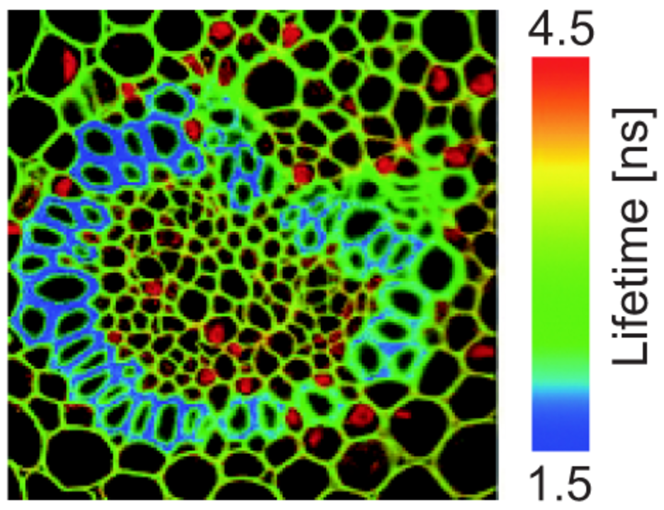
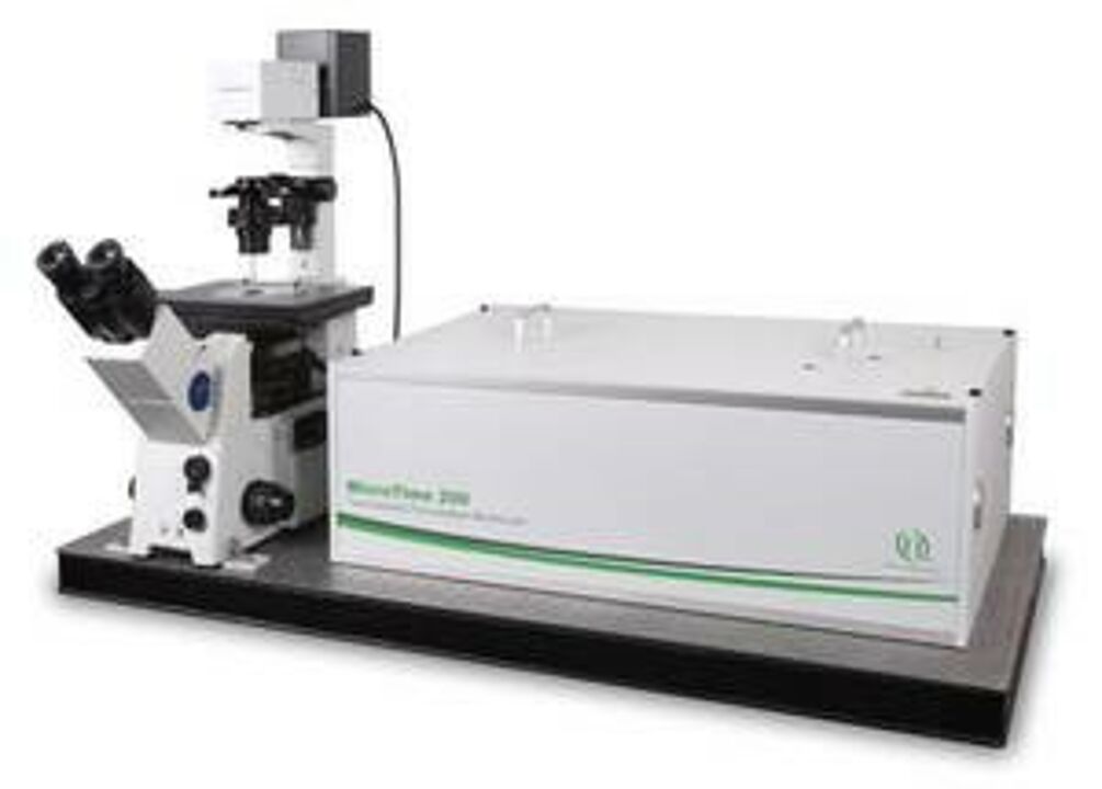
FLIM – Fluorescence Lifetime Imaging Microscopy
FLIM measures the time a fluorophore remains in an excited state before emitting a photon. This so-called fluorescence lifetime is a characteristic parameter of each fluorescent dye or molecule that may change with its nanoscopic surroundings or its conformational state. Lifetime information probes the molecular environment for its composition, such as ion concentration, pH, lipophilicity, or the binding to other molecules.
Lifetime information is independent of fluorophore concentration, so FLIM can be very useful for functional imaging in terms of investigating molecular function, interactions, and environment. Biosensors can also be exploited with FLIM to study cellular microenvironments. FLIM can be used to discriminate fluorescence probes that have overlapping emission spectra and eliminate unwanted background signals.
It also allows highly accurate FRET imaging, eliminating the crosstalk and/or bleaching problems inherent to standard ratio metric FRET imaging, and it provides an elegant tool for (time-resolved) anisotropy measurements.
EQUIPMENT
FLIM measures the time a fluorophore remains in an excited state before emitting a photon. This so-called fluorescence lifetime is a characteristic parameter of each fluorescent dye or molecule that may change with its nanoscopic surroundings or its conformational state. Lifetime information probes the molecular environment for its composition, such as ion concentration, pH, lipophilicity, or the binding to other molecules.
USER INFORMATION
In general, we offer four types of access to our shared research infrastructure:
- Research projects
- Full research services
- Trained user access | user labs
- Shared technology platform
Typically, these are set but not limited by the offered technology or instrument, and differ in the required user expertise, the usability of a technology, the user’s pre- and postprocessing input, and the underlying operational models.
Research project
Research projects are the equivalent of contract research organizations (CRO). The customer submits the starting material/sample and receives the ready-to-use data for publication. Hence, core facility members are often co-authoring and involved in the entire publication process.
Full research service
The user submits the sample, we perform a pre-defined workflow (incl. QC) and process the raw data. Data interpretation or contributions to publications cannot be offered in this service mode.
Trained user access | User labs
VBCF experts maintain an instrument park and train users to operate it. This requires a certain level of expertise, maturity of the offered technology, a hands-on attitude and reliability from the user.
Shared technology platform | Instrument park
These technologies require expert knowledge to run the offered instruments. Experts are hired by one of the research institutes on the Vienna BioCenter Campus. The experts maintain an instrument park, run the experiments and train other trainers. Machines can only be operated by experts.
The VBCF Light Microscopy facility provides trained user access.
In general, the VBCF General Cooperation Conditions apply. If you are interested in using the instrument, please contact the microscopy team.
We require acknowledgement of facility use in publications. A simple statement is sufficient and can be placed in the Materials and Methods section or in the Acknowledgments section, depending on the journal format.
Suggested format:
The XXXXXX was performed by the Light Microscopy Facility at Vienna BioCenter Core Facilities (VBCF) and the IMP/IMB/GMI BioOptics Facilty, members of the Vienna BioCenter (VBC), Austria.