


Immunohistochemical stains
The Histopathology facility offers chromogenic and fluorescent immunostaining for a variety of markers in diverse target tissues. Clicking on the links will take you to high resolution images corresponding to the representative examples listed below.
- Neuronal nuclear antigen (RBFOX3/NeuN)
- Glial fibrillary acidic protein (GFAP)
- Tyrosine hydroxylase (TH)
- Myelin basic protein (MBP)
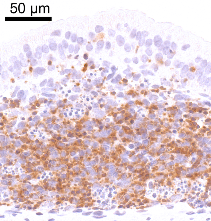
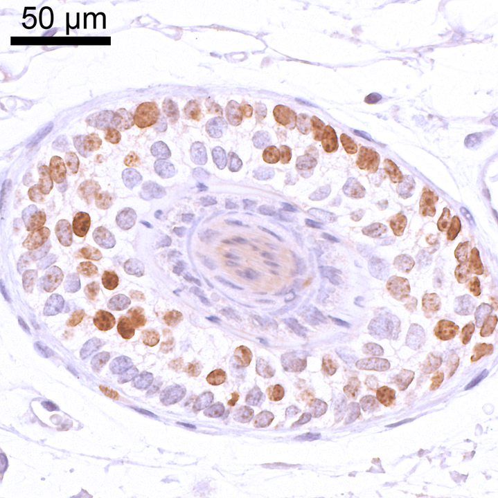
2. Immune / inflammatory cell markers
- Forkhead box protein 3 (FOXP3)
- CD3 antigen, epsilon (CD3e)
- Protein tyrosine phosphatase receptor type C (B220/CD45R)
- CD44 antigen (CD44)
- Adhesion G protein coupled receptor E1 (ADGRE1/F4.80)
3. Vascular and stromal markers
- Platelet endothelial cell adhesion molecule 1 (CD31)
- Collagen type IV (COL4) & Platelet endothelial cell adhesion molecule 1 (CD31]
- Caspase 3, cleaved (CASP3, cl)
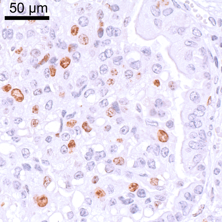
5. Cellular proliferation markers
- Antigen Ki-67 [Ki-67]
- Histone H3, phosphorylated (PHH3)
- Bromodeoxyuridine (BrdU)
6. Histones and DNA damage markers
- Histone 2A histone family, member X (gammaH2AX)
- Histone 2A histone family, member Y, variant 1 (mH2A1.1)
- Tumor protein 53 binding protein 1 (TP53BP1]
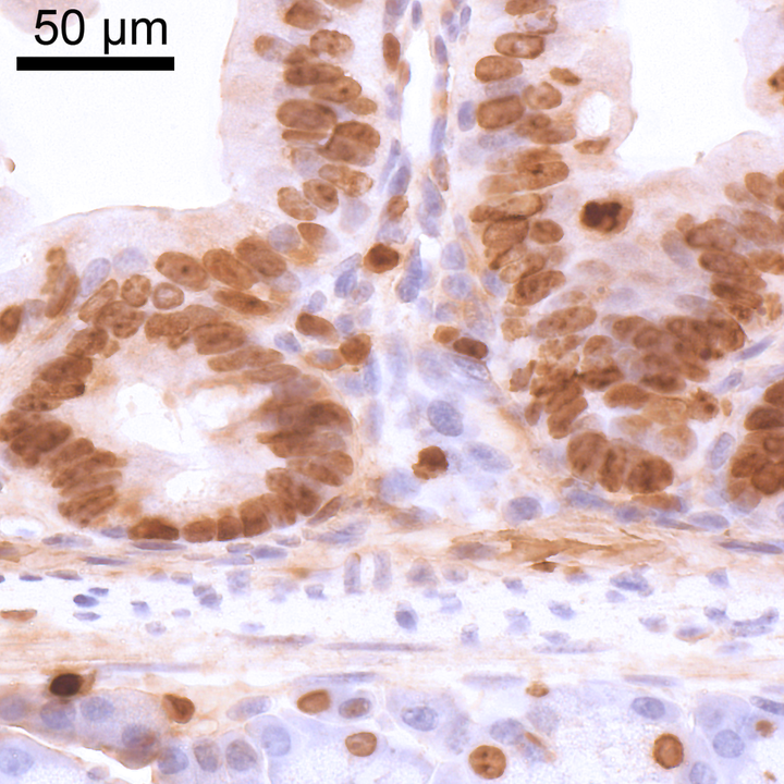
7. Epithelial, myoepithelial, basal, stem cell markers
- Sex determining region Y box 1 (SOX1)
- Transformation related protein 63 (TRP63)
- Epithelial cell adhesion molecule (EPCAM)
- Keratin 5 (KRT5)
- Keratin 19 (KRT19)
- Transformation related protein 63 (TRP63) & Keratin 14 (KRT14)
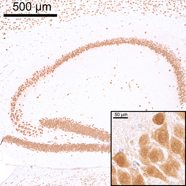
Neuronal nuclear antigen (NeuN)
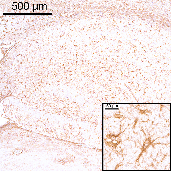
Glial fibrillary acidic protein (GFAP)
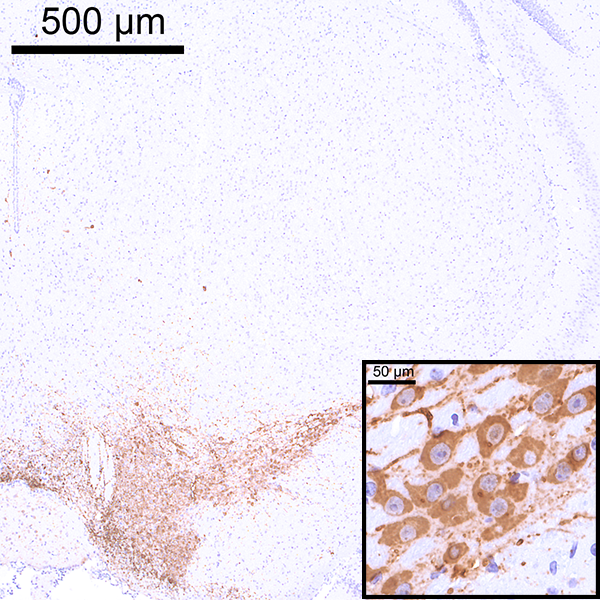
Tyrosine Hydroxylase (TH)
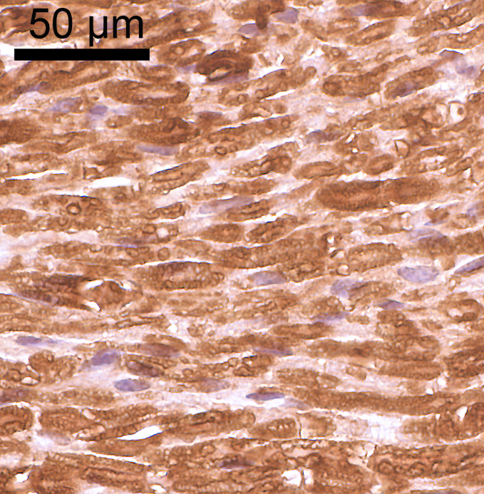
Myelin basic protein (MBP)
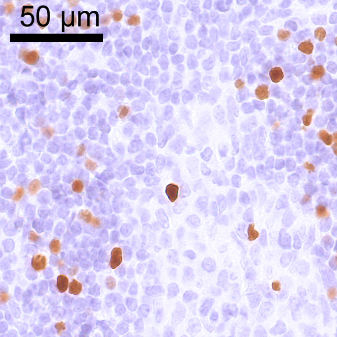
Forkhead box protein P3 (FOXP3)
CD3 antigen, epsilon (CD3e)
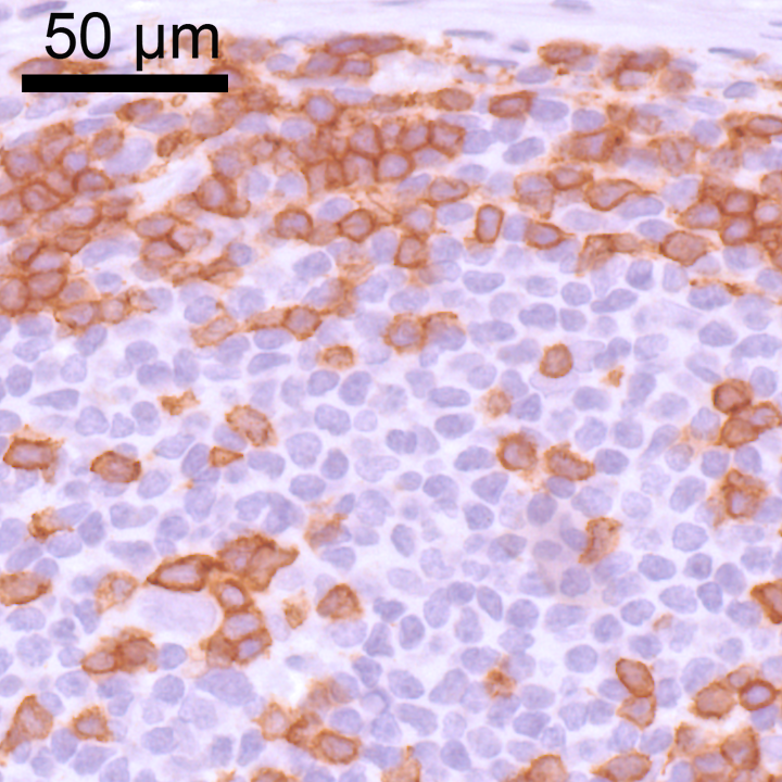
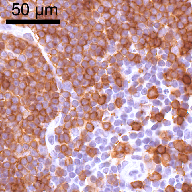
Protein tyrosine phosphatase receptor type C (B220/CD45R)
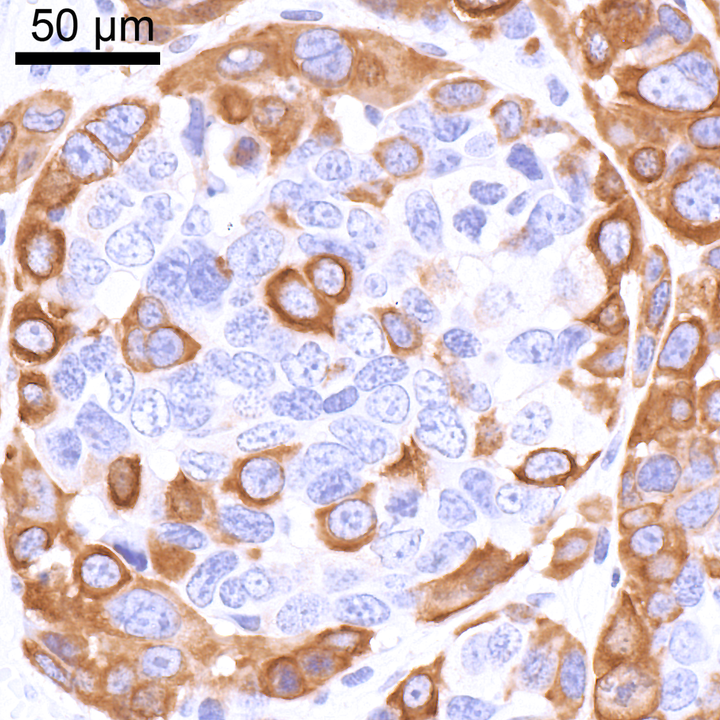
CD44 antigen (CD44)

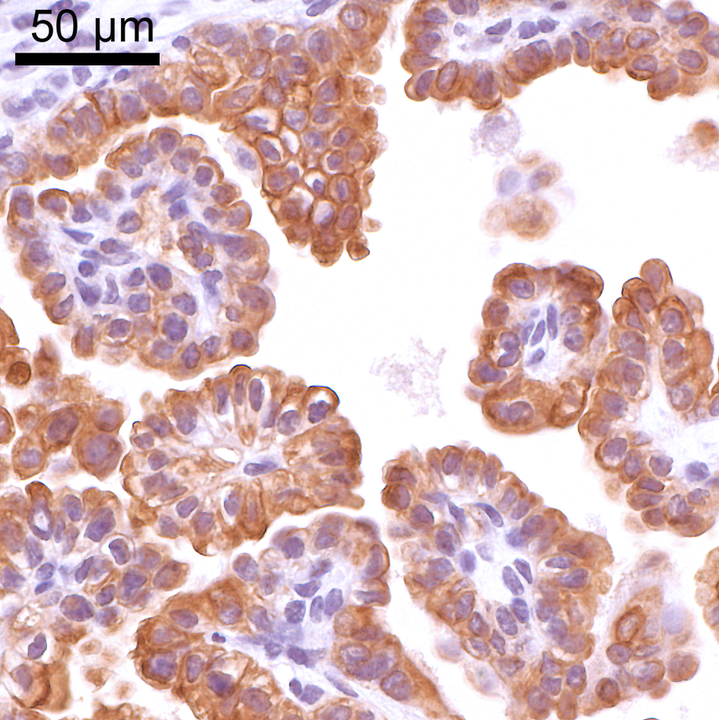
Adhesion G protein coupled receptor E1 (ADGRE1/F4.80)

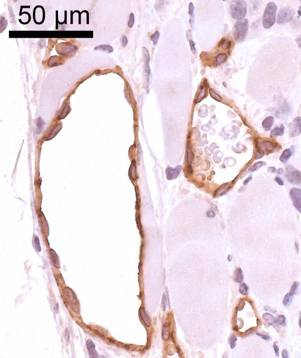
Platelet endothelial cell adhesion molecule 1 (CD31)
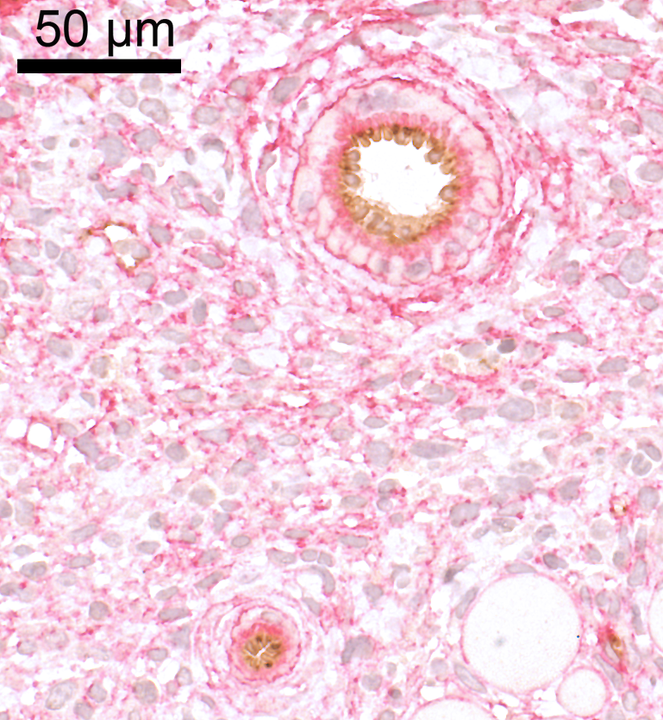
Collagen type IV (COL4) & Platelet endothelial cell adhesion molecule 1 (CD31)
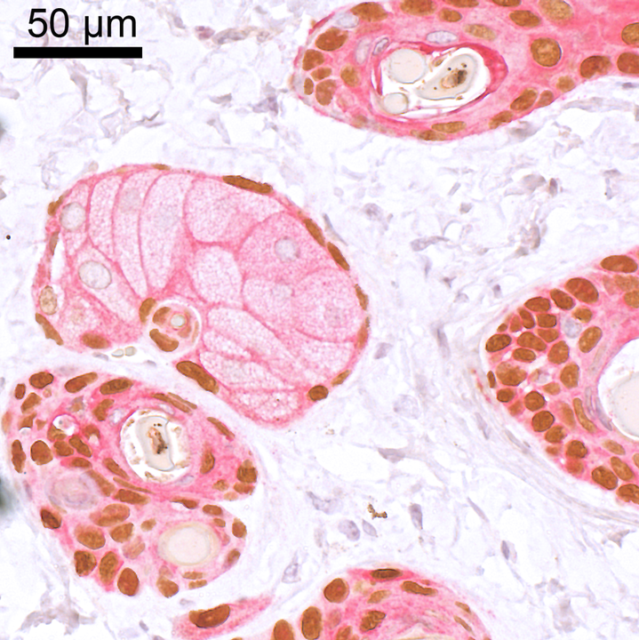
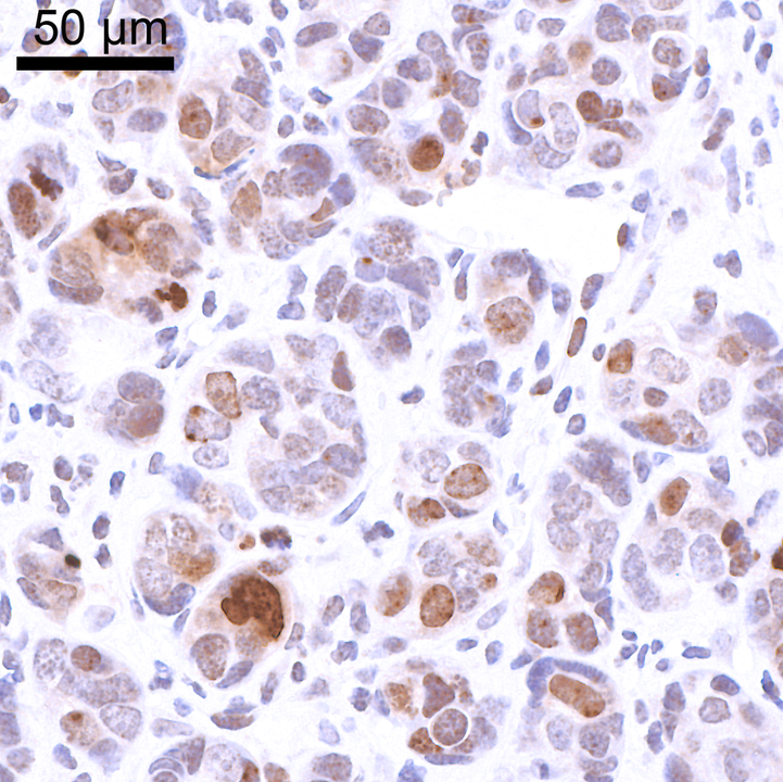
Caspase 3, cleaved (CASP3cl)
Caspase 3, cleaved (CASP3cl)
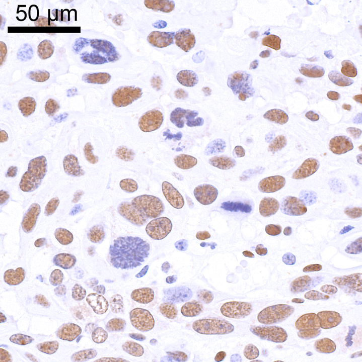
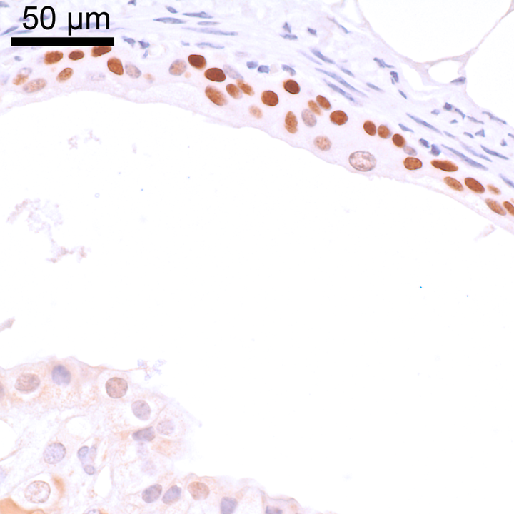
Antigen Ki-67 (Ki-67)
Antigen Ki-67 (Ki-67)
Histone H3, phosphorylated (PHH3)

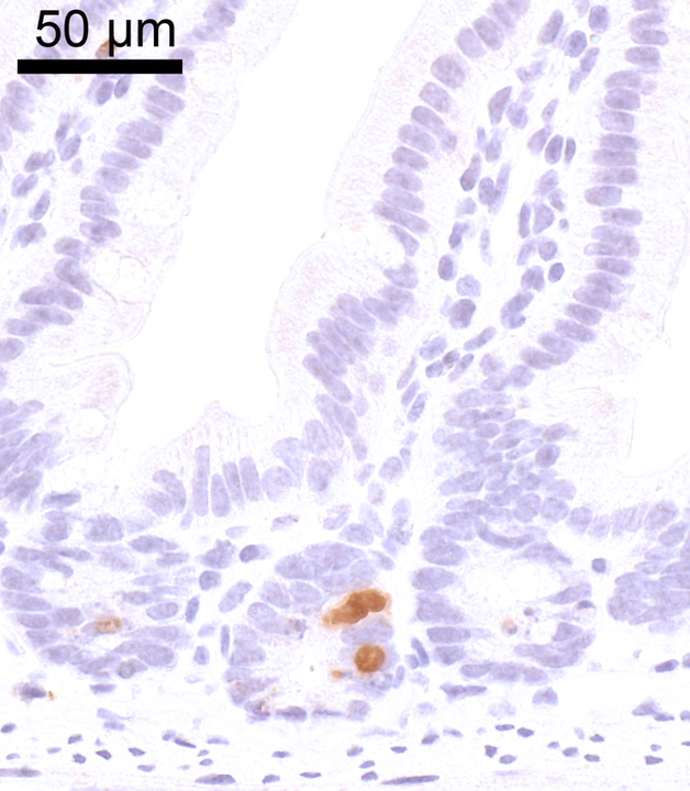
Bromodeoxyuridine (BrdU)
Histone 2A histone family, member X (gammaH2AX)
Histone 2A histone family, member Y, variant 1 (mH2A1.1)
Tumor protein 53 binding protein 1 (TP53BP1)
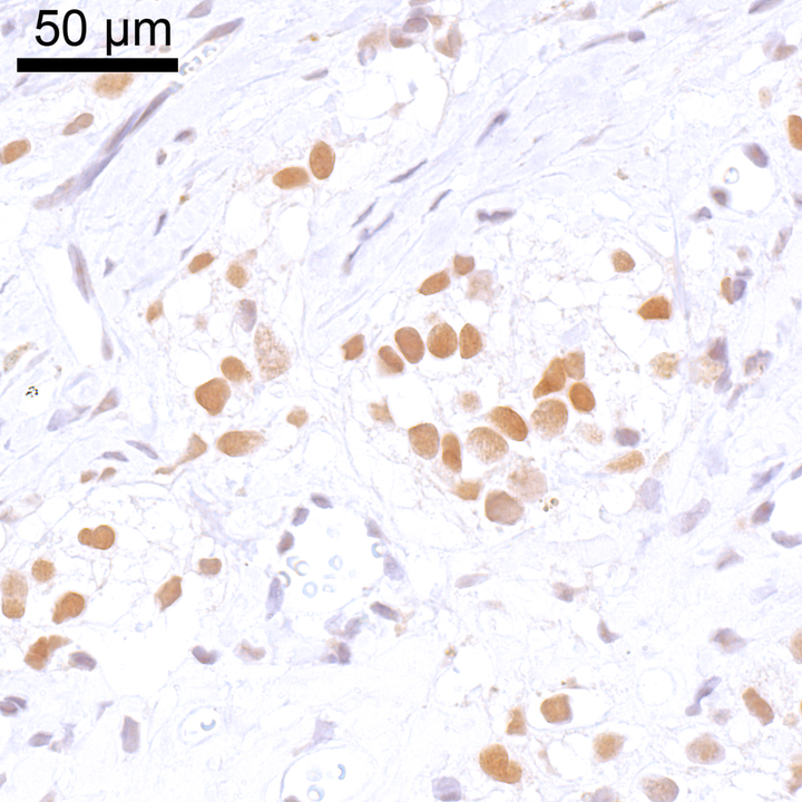
Sex determining region Y box 1 (SOX1)
Transformation related protein 63 (TRP63)
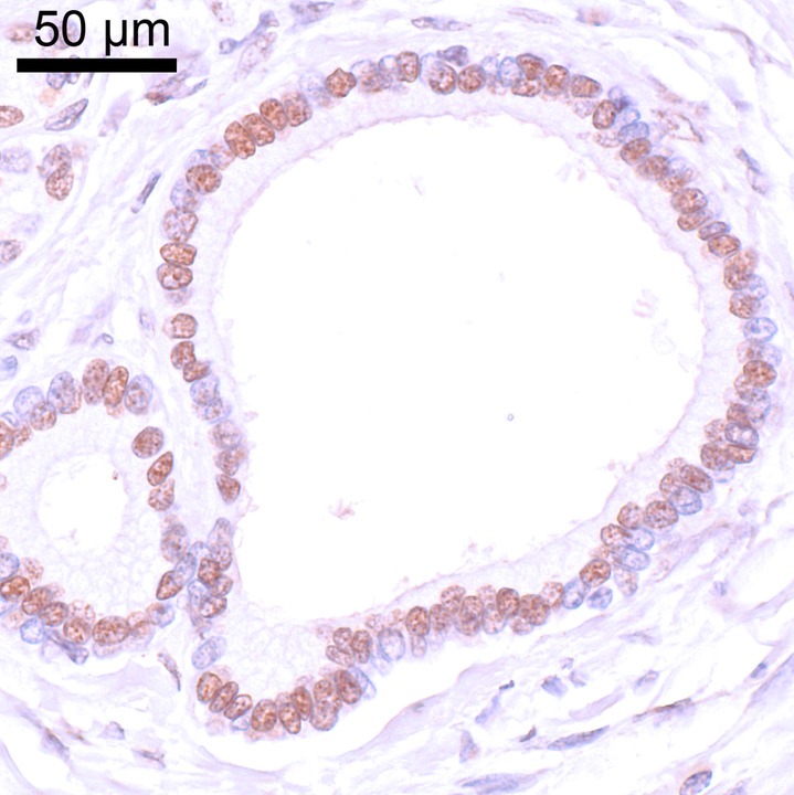
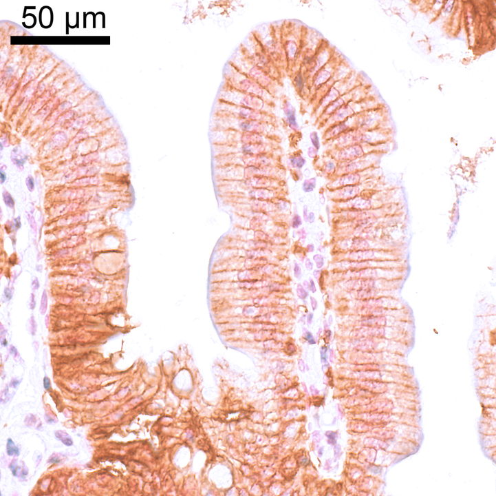
Epithelial cell adhesion molecule (EPCAM)
Keratin 5 (KRT5)
Keratin 19 (KRT19)
Transformation related protein 63 (TRP63) & Keratin 14 (KRT14)